Heart & Vascular Transcript
Heart & Vascular – Leo Polosajian, MD Video
Announcer: Electrophysiology is a highly specialized type of cardiology, which focuses on diagnosis and correction of the pacing of the heart, its electrical impulses and arrhythmias.
Visuals: Announcer is a male, wearing a suit. He is in front of a news studio background with glass and wood panels and a large monitor with the title “Heart Arrhythmias” displaying a motion graphic of a beating heart; heart rate monitor displaying heart rhythm numbers including 65, 75, 88, 71, and 136.
Announcer (V.O.): We went to Valley Presbyterian Hospital in the San Fernando Valley and spoke to Dr. Leo Polosajian, a cardiac rhythm specialist and asked him about his specialty, electrophysiology.
V.O. visuals: Main exterior entrance of Valley Presbyterian Hospital; Dr. Polosajian speaking with a senior male patient.
Leo Polosajian, MD: Electrophysiology specifically is to study the electrical system of the heart. So, for instance, when someone passes out, or for example, when someone has an extremely rapid heart rate, or palpitations, shortness of breath because of palpitations, per se, or dizziness, lightheadedness, syncope, passing out…
V.O. visual: Dr. Polosajian, wearing a suit and tie, is being interviewed in a studio in front of a brightly colored abstract background. He is introduced with the title “Leo Polosajian, MD – Valley Presbyterian Hospital” in the lower-third.
Polosajian (V.O.): …then we basically need to look at their electrical system.
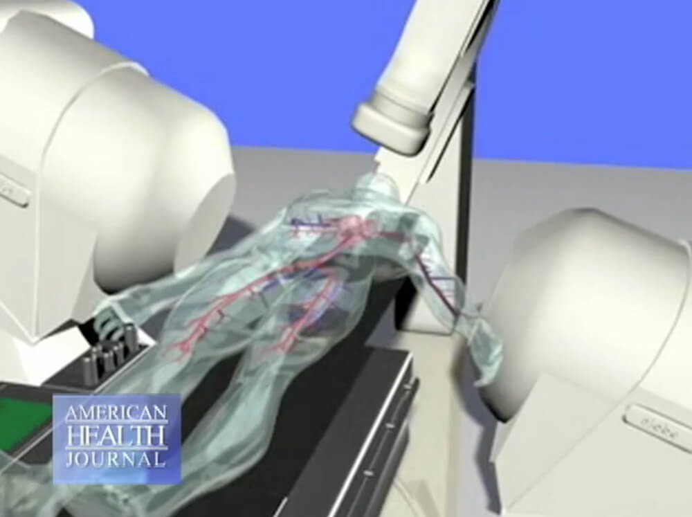
V.O. visual: Motion graphic diagram of a machine scanning a human with a heart and arteries on view (images below).
Announcer (V.O.): Dr. Polosajian talks about atrial fibrillation or AFib.
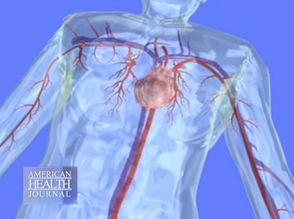
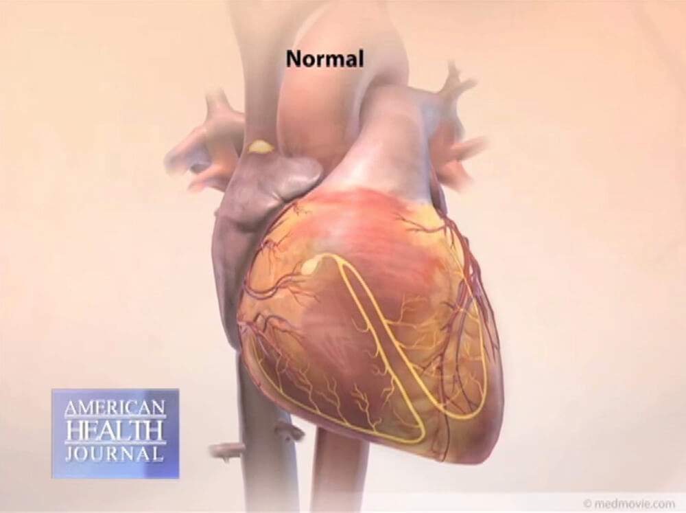
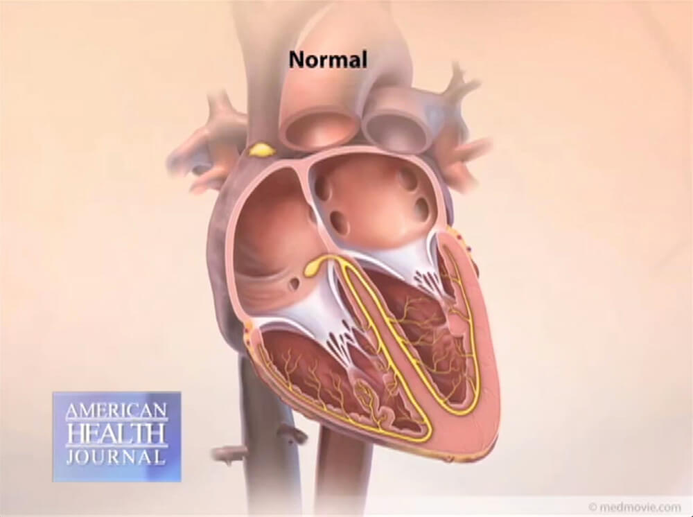
V.O. visuals: Close-up of heart and arteries diagram; motion graphic of a heart beating normally with the title “Normal" (images below).
Polosajian (V.O.): AFib is the most common arrhythmia. AFib is when the upper chamber of the heart starts to fibrillate, starts to quiver. And one risk factor of that is…
V.O. visuals: Motion graphic of heart beating irregularly with the title “Atrial Fibrillation,” followed by a description “Disorganized atrial signals” indicating an area of the diagram where the heart is quivering (images below).
.jpg)
.jpg)
Polosajian: …if you get a stroke, in the event that you’re in AFib for longer than 48 hours, constantly. Sometimes AFib can present itself…
Polosajian (V.O.): …as paroxysmal, it comes and goes, then it may be persistent, then it may be chronic.
V.O. visual: Polosajian in his office talking on the phone.
Polosajian: This is the pulmonary artery. This goes toward the lung, this vessel. This vessel is connected to the right side of the heart, the pulmonary because it takes the oxygen, the blood, brings it to the lungs in order for the blood to get oxygenated, then from there it turns from the lungs and comes through the pulmonary vein. From the pulmonary vein it enters into the left atrium, from the left atrium into the left ventricle, from the left ventricle into the aorta, and the aorta to the body. So the reason this pulmonary vein is important is because when you have atrial fibrillation, and especially if it is paroxysmal, or persistent, many times, the majority of the time, it starts from the pulmonary vein and the electricity starts from there and it shoots into the left atrium, from the left atrium into the right atrium and arrhythmia continues.
V.O. visuals: Polosajian in studio holding up a 3rd model of a heart, pointing at various parts of the heart as he describes it in the audio. Camera zooms in for a close-up view. When he talks about atrial fibrillation, the title “Dr. Polosajian discusses Atrial Fibrillation” appears in lower-third.
Polasajian (V.O.): AFib, in the past, we could not cure it. (inaudible) Then surgical procedures came about where they did basically a maze procedure. They actually had to do an open heart surgery and take the left atrium…
V.O. visuals: Healthcare worker pulling a paper with a patient’s test results from an EKG machine; close-up of page with results; surgery team working in operating room working on a patient; close-up of a maze procedure; motion graphic of white lines being drawn on the atrium area of the heart.
Polasajian: … cut it in pieces, resuture it together, kind of like make a maze out of this. And when they did that, they discovered that the electrical system could not fibrillate any longer and sinus rhythm, which is a normal rhythm, took over.
Polasajian (V.O.): Nowadays what we do is ablation. Same technique in a way but without open heart surgery. The procedure is done as an outpatient. The patient comes and gets it done in approximately three hours and stays one night, overnight for observation, goes home the next day. Recovery is so fast they go back to work in three days or two days.
V.O. visuals: Motion graphic of the inside of the body in the heart area with an ablation tube; motion graphic close- up of an ablation tube near the opening of the skin (images below):
.jpg)
.jpg)
(V.O. visuals continued) healthcare worker preparing the cath lab room; close-up of the equipment dashboard; close-up of the computer monitor screen with test results; close-up of monitor with heart rate.
Polasajian: This is a pacemaker. As you can see it is very small.
V.O. visual: Polasajian holds up a small oval device, about 2 inches diameter and about 1/4 inch in depth.
Polasajian (V.O.): Pacemakers are implanted to prevent slow heart rates.
V.O. visuals: A motion graphic, “Courtesy of the American Heart Association” shows a heart with wires extended into it from a pacemaker that is attached to the inside of the chest near the shoulder. The graphic starts with the title “Sensing” with the description “The pacemaker is a device that can sense this abnormal beat when it occurs.” The heart is beating slowly and an inset of a “Sick Sinus ECG” displays a slow rate. The title then changes to “Pacing” with the description “The pacemaker corrects the abnormal heartbeat by firing electrical signals to restore a normal heart beat.” The heart is beating at a normal speed and an inset of a “Normal ECG” displays a normal rate (images below).
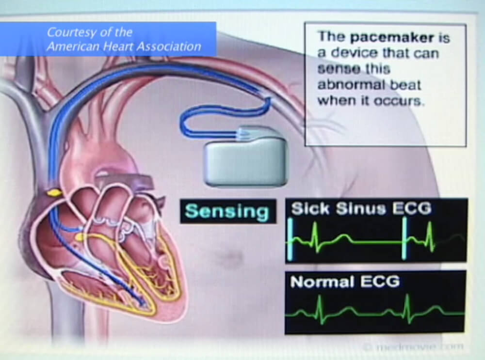
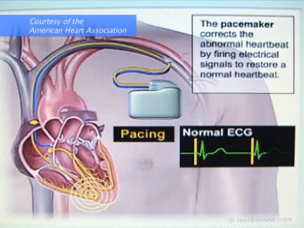
Polasajian: Not rhythm. Rates. So when the heart rate goes down, for example, at less than sixty beats per minute and the pacemaker is set at sixty, the pacemaker is a demand pacemaker. It will kick in, in order to restart the heart, per se, electrically, to pace the heart.
Annoucer (V.O.): The doctor tells us what he sees for the future in treating heart arrhythmias.
V.O. visuals: Polasajian explains information on a monitor to his senior male patient; monitor next to the cath lab patient room with heart scan, rate and other detailed information.
Polasajian (V.O.): I believe that ablation will stay because it is helping a lot of people fairly quickly. Outpatient procedures. I believe medications may grow, specifically for arrhythmias but if…
V.O. visuals: Camera pans the cath lab patient room to reveal a bed, equipment and monitors; close-up of the monitor showing a heart graphic with arrows moving in a circular motion; female healthcare worker sitting in front of a monitor reviewing the data.
Polasajian: …you catch the arrhythmia early on you can definitely help that patient and prevent many other symptoms from coming.
Visual: Polasajian being interviewed in the studio.
End screen is black.