Stem Cells / Orthopedics Transcript
Stem Cells / Orthopedics – Eric Ferkel, MD Video
Opening screen with title “American Health Journal” and swirling graphics in motion over blurred images of physicians.
[Serious music]
Announcer (V.O.): All types of sports have a potential for injury, whether from trauma, contact with other players or overuse. Orthopedic surgeon Dr. Eric Ferkel at Valley Presbyterian Hospital discusses the newest stem cell techniques to help these injuries heal faster. He defines what a stem cell is and how the procedure promotes healing.
V.O. visuals: Men in a gym playing a fast game of basketball; men on a team playing competitive soccer on a field; young men in the starting position on an outdoor track ready to race; Dr. Erik Ferkel being interview in a studio; surgery team working on a patient in the operating room; close-up of doctor looking into a microscope.
[Music fades out]
Eric Ferkel, M.D.: A stem cell is any cell in your body that essentially can become and differentiate into a different type of cell…
Visual: Close-up of Ferkel in a suit and tie being interviewed in the studio against a bright blue and black background. He is introduced with the title “Eric Ferkel, MD – Valley Presbyterian Hospital” in the lower-third.
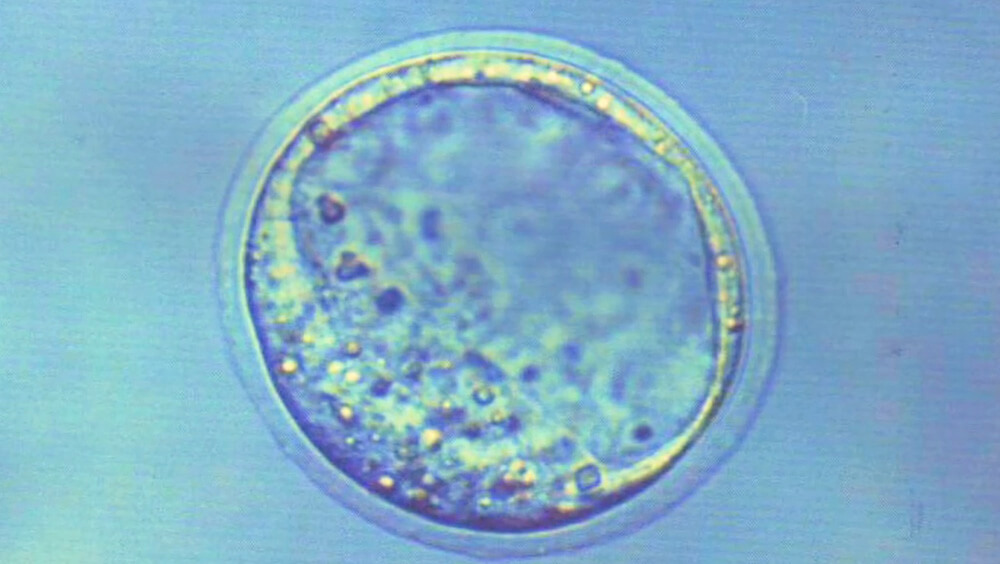
Ferkel (V.O.): …depending on what the cell’s lineage is or its background is. So for example, if we put a stem cell that we take out of your blood, for example, or we take out of your bone marrow…
V.O. visual: Close-up of cell under a microscope (image below).
(V.O. visuals continued): spinning machine used in stem cell extraction; plastic tube with blood flowing through it; another view of the machine with the tube going into the top of it; motion graphic of blood cells falling from the top of frame (image below).
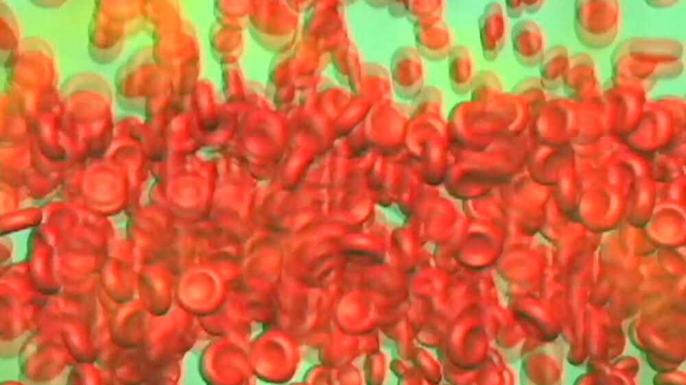
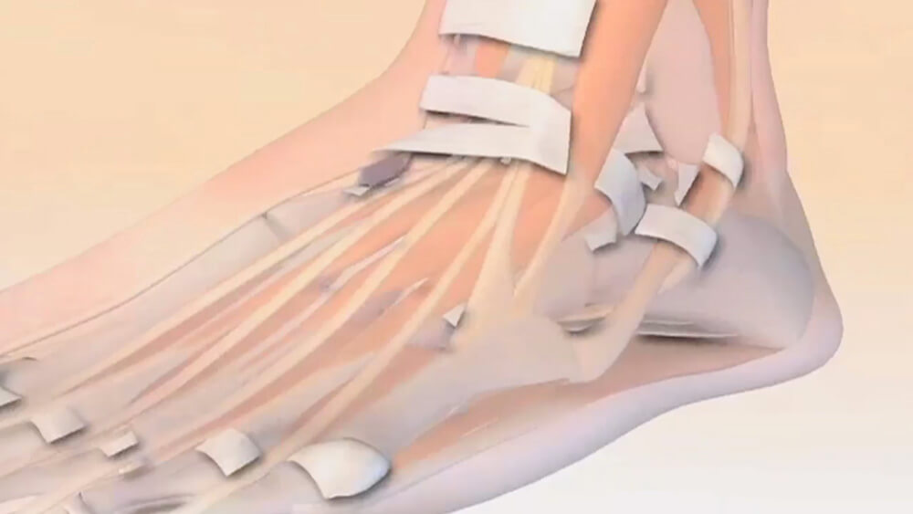
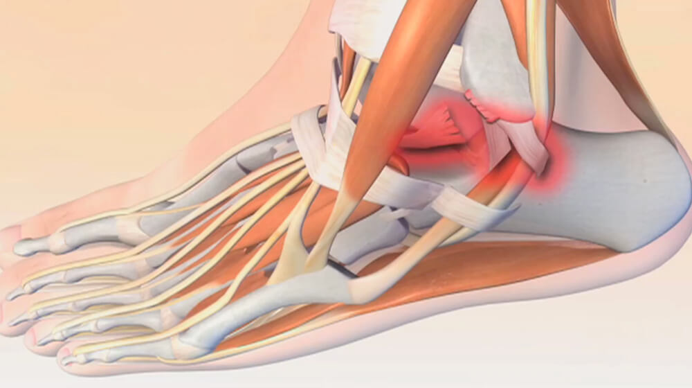
Ferkel: …and we put the bone marrow stem cells into your ankle, for example, the idea is to help bring new nutrition to the ankle where the cartilage injury would be…
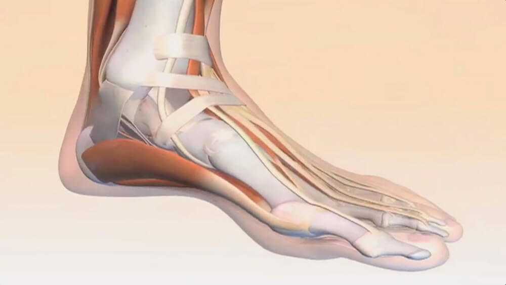
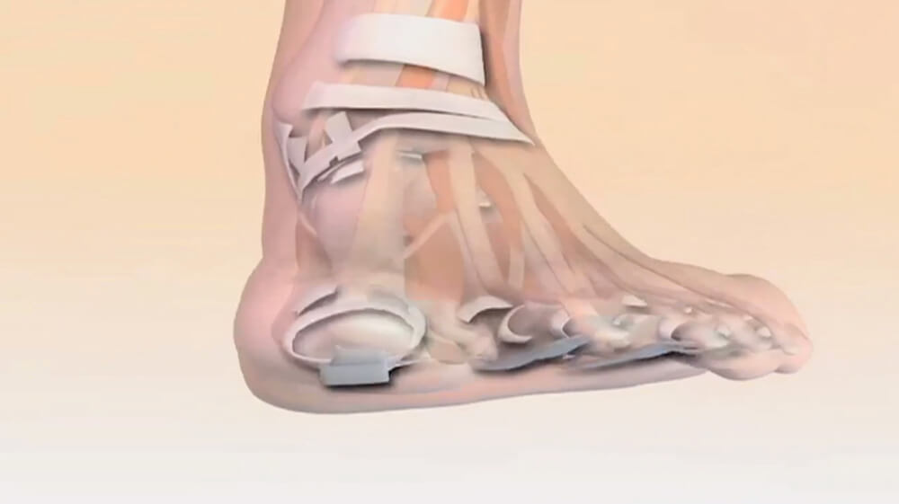
Ferkel (V.O.): …in bringing new growth factors there. So these cells can turn into growth factors and provide new healing and different forms of nutrition to help increase and expedite the healing process.
V.O. visual: Motion graphic moving up and down a foot and interior ankle showing bones and cartilage, then spins to show front of foot and toes, then spins again to show the side of foot and outer ankle area with ligaments shaded in red (images below).
Announcer (V.O.): Dr. Ferkel describes how an injury is evaluated to determine if stem cell therapy is the best option.
V.O. visual: A doctor points to an X-ray of foot that shows pins that have been placed in it.
Ferkel: First, when someone comes in with an ankle injury and we obtain imaging studies such as an X-ray, then followed by an MRI and or a CT scan, and we identify possibly a cartilage…
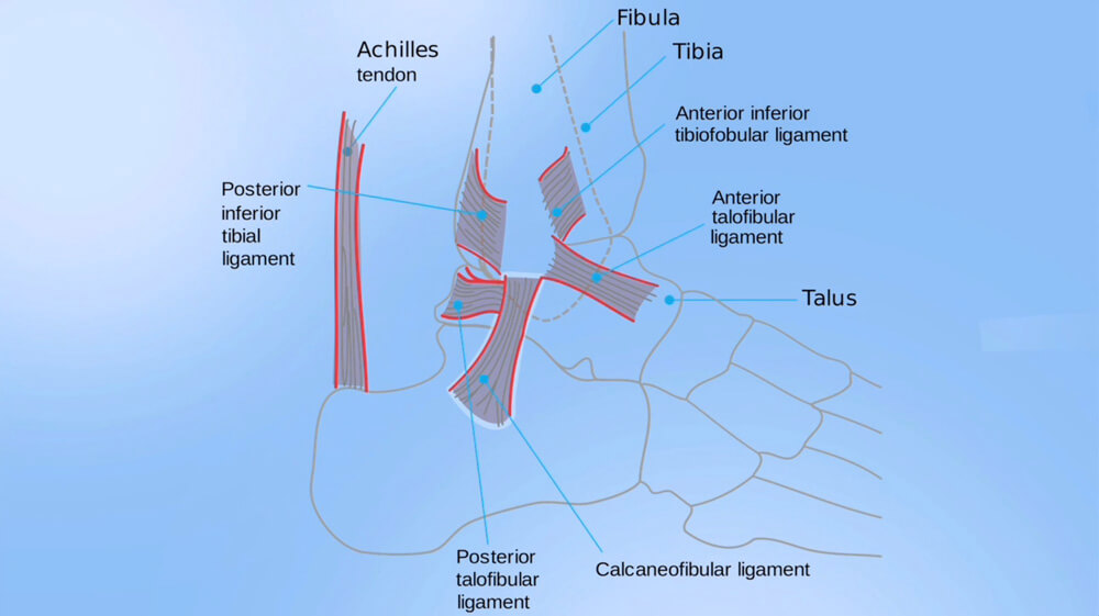
Ferkel (V.O.): …injury in the ankles, specifically perhaps on the talus, which is the bone that makes up the bottom portion of your ankle, or on the tibia, which makes up the top portion of your ankle.
V.O. visuals: Close-up of a doctor holding a 3-D model of a foot and ankle and pointing to talus area of ankle; graphic illustration of the foot and ankle with labels: Achilles tendon (running vertically in the back of the ankle area), Posterior inferior tibial ligament (located in the top interior of the ankle toward the back), Posterior talofibular ligament (located under the posterior inferior tibial ligament), Calcaneofibular ligament (located under the posterior talofibular ligament), Fibula (the bone located above the ankle), Tibia (located under the Fibula), Anterior inferior tibiofobular ligament (located in the top interior of the ankle toward the front), Anterior talofibular ligament (located under the anterior inferior tibiofobular ligament), and Talus located under the Anterior talofibular ligament) (image below):
Ferkel: Based off the size of the cartilage injury, we then determine well, what are the options that this patient has available to them to help improve the chances that this will heal…
Ferkel (V.O.): …and get better and have them improve their functionality, getting back to playing the sports they want to be doing, or just walking and living a pain-free life.
V.O. visuals: Young woman practicing soccer by bouncing the ball upward with her knees; another clip of her walking and holding the ball.
Ferkel: So based off of the size of the lesion, your algorithm could begin with basically going into your ankle…
Ferkel (V.O.): …with an orthoscope, which is a little video camera that you put inside the ankle joint and you look around, and you find the lesion. And, you look at the lesion and you determine ‘ok this is an amenable to repair,’ meaning that we would usually scrape out where the tear is in..
V.O. visuals: Surgical team working on a patient in the operating room; monitor displaying the inside of patient’s ankle
Ferkel: …the cartilage and make little poke holes into the bone to help increase stem cells from in your own bone and…
Ferkel (V.O.): …improve the milieu, which is kind of the environment that the cartilage is growing in. Secondly, then we would take bone marrow aspirate. So we take aspiration from your bone marrow and put it down into a centrifuge and get the concentrated amount of bone marrow aspirate. Then after the surgery is done we would then inject it into your ankle. Studies have shown that using bone marrow aspirate concentrate or something called platelet rich plasma, and the jury’s still out on which is better, or…
V.O. visuals: Close-up of team working on the arm of a patient to remove bone marrow; technician placing bone marrow vial on top of aspirate device; technician placing vial inside and closing the lid; two female lab workers sitting at a desk conducting tests; close-up of technician placing tube vial inside an empty vial.
Ferkel: …if they’re good in combination, injecting them into your ankle can help increase the healing of that cartilage injury that you had.
Announcer (V.O.): Dr. Ferkel gives us a case study of a recovery from injury using stem cell treatment.
V.O. visual: Ferkel walking up the hospital’s main hallway toward and past the camera.
Ferkel: I had a twenty-three year old male who came in and saw me. He likes to play beach volleyball on the weekends…
Ferkel (V.O.): …here in Los Angeles. Given his activities being in volleyball, he jumps a lot when he plays volleyball on the beach…
V.O. visual: Young men on the beach playing volleyball.
Ferkel: …especially. And, he came in with pain along his Achilles tendon and we worked him up, started him doing physical therapy initially. It was getting a little better, not great. And, got an MRI and showed that he had tendinopathy, meaning that the tendon was inflamed, wasn’t torn to a significant extent that you’d want to do surgery on…
Ferkel (V.O.): …and given that he was a young male, wanting to get back to playing beach volleyball - it was the summertime – we decided let’s try to do platelet rich plasma for his Achilles tendon. But…
V.O. visuals: Various close-ups of lab technician inserting a substance into an empty vial; two technicians at a table working with the vials.
Ferkel: …I think for certain patients, this is a young, healthy, active male, it’s worth a shot, no pun intended, but it’s worth a shot to take his own platelet rich plasma…
Ferkel (V.O.): …which is taking the blood from his arm, spinning it down, extracting the stem cells and then injecting it into his Achilles tendon, at the area where the injury was.
V.O. visuals: Technician placing vials in an aspirate device; close-up of female doctor holding up vial of stem cells.
Ferkel: So I saw him initially three weeks later and he was telling me how his pain has essentially not gone away one hundred percent but its gone away about eighty percent.
Ferkel (V.O.): And then again, three weeks after that, we had him back out playing volleyball right now. It’s very rewarding to see these athletes and people who we are taking care of, who are just your regular weekend warriors, getting back out and doing the sport they like to be doing.
V.O. visual: Men playing volleyball on the beach; young men on outdoor race track in starting position and taking off up the track towards and past the camera; close-up of feet and ankles of men playing basketball inside a gym; camera pulls back to show them jumping up to make and block a basket.
Ferkel: There’s so much potential in doing either cartilage treatment or tendinopathies with stem cells that it’s definitely…
Visual: Ferkel being interviewed in the studio.
Ferkel (V.O.): …a great opportunity for treating somebody who needs it.
V.O. visual: Teenage boy and girl outside playing basketball on a park court.
End screen is black.